A student was given a sample of two tissues. He observes the tissues under the microscope and draws their figures (1 and 2) as shown below .
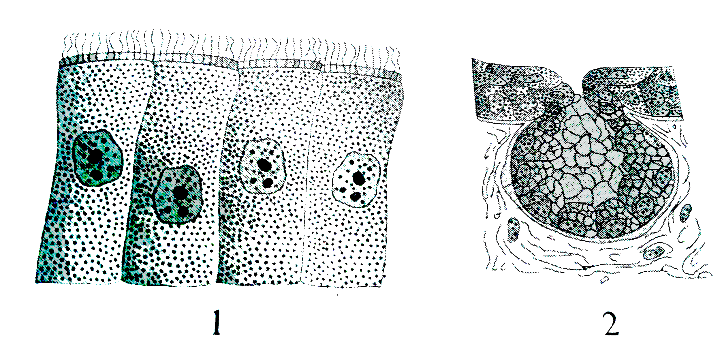
Identify the tissues (1 and 2)
1:Columnar cells bearing cilia , 2:Unicellular glandular epithelium
1:Cuboidal cells bearing cilia , 2:Multicellular glandular epithelium
1:Compound cells bearing cilia , 2:Unicellular glandular epithelium
1:Columnar cells bearing cilia , 2 : Multicellular glandular epithelium
The correct Answer is:D
Step by step video solution for A student was given a sample of two tissues. He observes the tissues under the microscope and draws their figures (1 and 2) as shown below . Identify the tissues (1 and 2) by Biology experts to help you in doubts & scoring excellent marks in Class 12 exams.
Updated on:21/07/2023टॉपर्स ने हल किए ये सवाल
समान प्रश्न
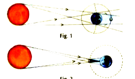
Name the type of eclipses shown in the Figures 1 and 2.
View SolutionA student was given a sample of tissue. He observes and concludes the following characters.
(i) The following The cells are composed of a single layer of tall and slender cells.
(ii) Their nuclei are located at the base.
(iii) Free surface may have microvilli .
(iv) It is found in the lining of stomach and intestines
(v) It help in secretion and absorption.
Based on the above features identify the epithelium.
View Solution
ज्ञान जांच
In the given flow chart the flow of water is shown from soil to xylem of the root. Identify the tissues involved in steps A nad B
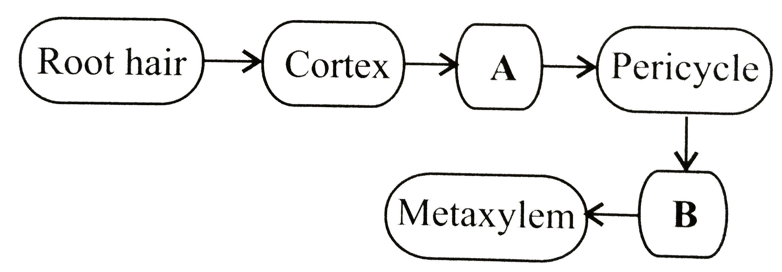 AA -Hypodermis , B - protoxylemBA - Medullary rays ,B - PhloemCA - Endodermis , B - PhloemDA - "Endodermis", B -"Protoxylem"
The given figure shows the diagrammatic represtation of exchange of gases at the alveolus and the body tissues with blood and trasport of oxygen and carbon dioxide. Identify the blood vessles A to D.
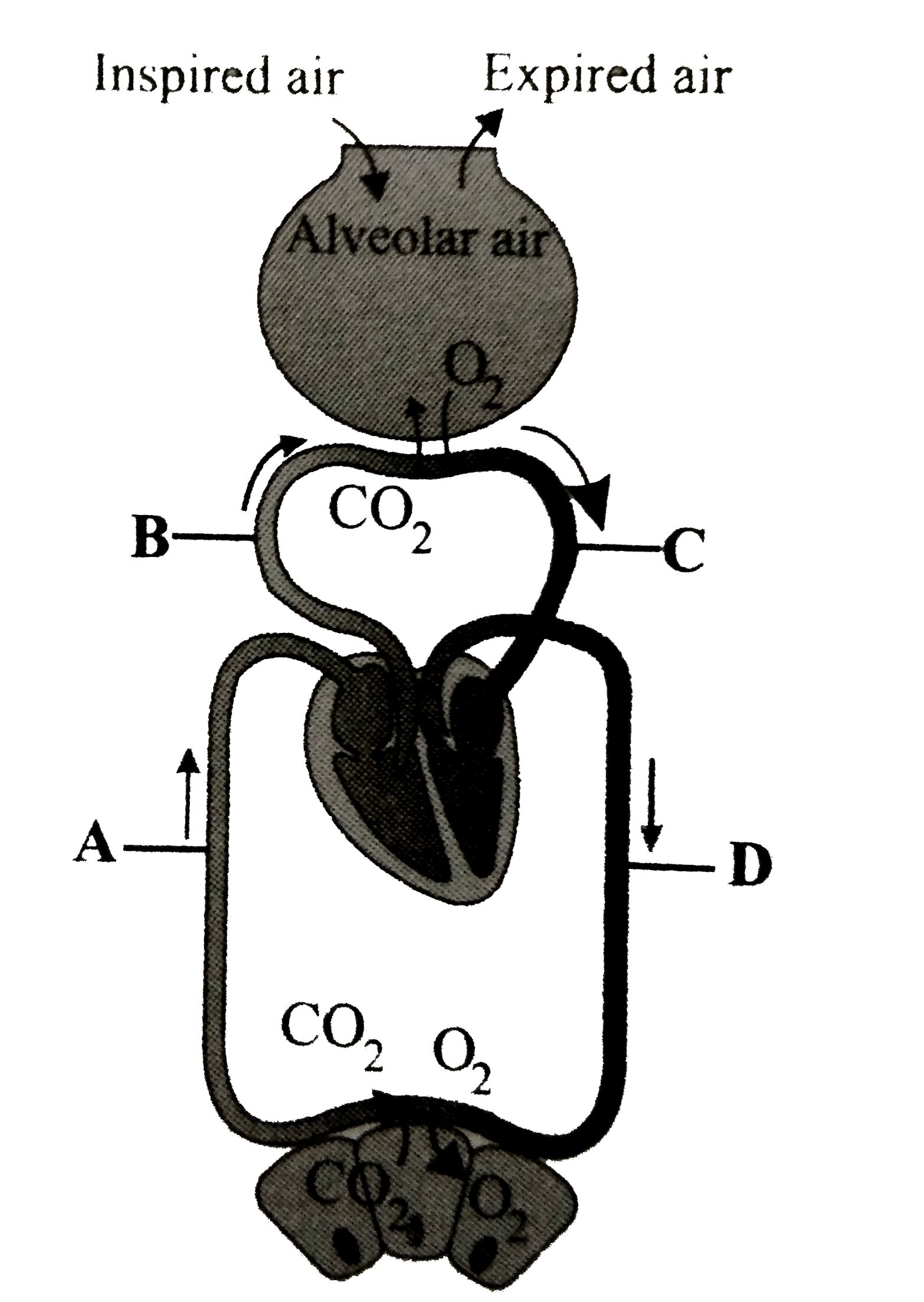 AABCDSystemic veinPulmonary arteryPulmonary veinSystemic arteryBABCDSystemic arteryPulmonary arteryPulmonary veinSystemic veinCABCDPulmonary arterySystemic veinPulmonary veinSystemic veinDABCDSystemic veinPulmonary veinPulmonary arterySystemic artery
The diagram given below shows the supportive connective tissue.

Identify the tissues A and B.
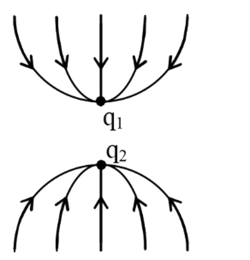
View SolutionIn the adjoining figure, the electric field lines for charges q1andq2 are shown. Identify the sign of the charges.
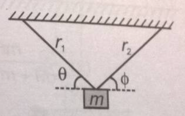
View SolutionA block is suspended by two light ropes r1andr2 as shown in figure . Then what is the tension the rope r2
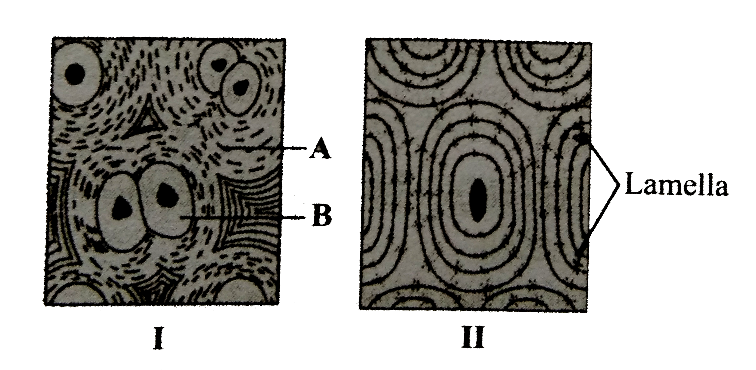
View SolutionGiven figures (IandII) show two specialised connective tissues. Identify the figures I,II and the parts labelled as A and B
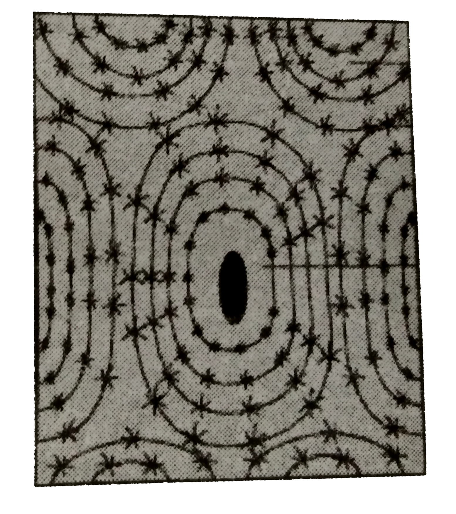
View SolutionIdentify the given below tissue with its type and select the right option for the two together
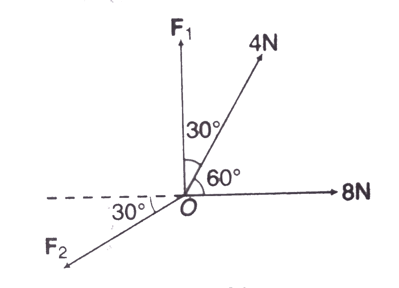
View SolutionAn object is in equilibrium under four concurrent forces in the direction shown in figure. Find the magnitudes of F1 and F2
View Solution

